Lupine Publishers Journal of Surgery and Journal of Case Studies: Currently case studies drag the concentration of the investigators since each case present provides deep understanding in diagnosis and treatment methods. It is devoted to publishing case series and case reports. Articles must be genuine
Tuesday, September 29, 2020
Lupine Publishers: Lupine Publishers | Allele Mining for the Reported...
Friday, September 25, 2020
Lupine Publishers: Lupine Publishers | Evaluation of Combinatorial Ca...
Lupine Publishers: Lupine Publishers: Lupine Publishers | Intervariet...
Tuesday, September 22, 2020
Lupine Publishers: Lupine Publishers | Perceived Effects of Resource-...
Lupine Publishers: Lupine Publishers | Intervarietal Hybridization an...
Friday, September 18, 2020
Lupinepublishers|OSMF-A Review
Lupine Publishers | Journal of Health Research and Reviews
Abstract
Oral submucous fibrosis is a premalignant condition that has received considerable attention in the recent past because of its chronic debilitating and resistant nature. In 600 B.C, Sushruta a well-known expert on Indian Medicine described in his classification of mouth and throat maladies mentioned about a condition (VIDARI) the features of which were progressive narrowing of mouth, depigmentation of oral mucosa and pain on taking food. The present article discusses etiopathogenesis, clinical features and medical management of this disease entity.
Keywords: OSMF; Pathogenesis; Review
Introduction
Oral submucous fibrosis is a premalignant condition that has received considerable attention in the recent past because of its chronic debilitating and resistant nature. It is now strongly believed that there is a definite relation of the condition with the habit of areca nut chewing [1]. Areca nut has been deeply rooted in Indian culture and been used as a mouth-freshening agent that has had various symbolic roles throughout Indian history [2]. The potential for malignant transformation in submucous fibrosis is considered high, and the disease affects persons of all ages and both sexes across the Indian subcontinent. Perhaps what is most disturbing about the condition is that it affects a number of adolescents as well. The disease occurs mainly in Indians. It affects between 0.2%-1.2% of urban population attending dental clinics in India. Pindborg JJ [3] defined oral submucous fibrosis an insidious, chronic condition that can affect any part of the oral cavity and sometimes even the pharynx. Although occasionally preceded by and/or associated with vesicle formation, OSF is always associated with juxta-epithelial inflammatory reaction followed by fibroelastic changes to the lamina propria with epithelial atrophy leading to stiffness of oral mucosa and causing trismus and inability to eat [3]. Rajendranelaborated Submucous fibrosis is an insidious, chronic disease affecting any part of the oral cavity and sometimes the pharynx. Occasionally it is preceded by and/or associated with vesicle formation and is always associated with a juxta-epithelial inflammatory reaction followed by progressive hyalinization of the lamina propria. The later subepithelial and submucosal myofibrosis leads to stiffness of the oral mucosa and deeper tissues with progressive limitation in opening of the mouth and protrusion of the tongue, thus causing difficulty in eating, swallowing and phonation [4].
Malignant Potential and OSMF
The precancerous nature of OSMF was first postulated by PAYMASTER in 1956, who described the development of a slow growing squamous cell carcinoma in one third of OSMF cases seen in Tata Memorial Hospital Bombay [5]. Epithelial dysplasia in OSF tissues appeared to vary from 7 to 26% depending on the study population .However, according to the current awareness of the disease and some refined criteria for grading dysplasia, it is reasonable to assume that the prevalence of dysplasia is more towards the midway of the reported range. Malignant transformation rate, of OSF was found to be in the range of 7-13% [1]. The hypothesis that dense fibrosis and less vascularity of the corium, in the presence of an altered cytokine activity creates a unique environment for carcinogens from both tobacco and areca nut to act on the epithelium is widely being accepted.
Some Common Classifications of OSMF
Khanna and Andrade [1] categorized OSF into different stages, as follows.
I. Group I: Very early
a. Normal mouth opening
b. burning sensation
c. Excessive salivation
d. Acute ulceration and recurrent stomatitis
II. Group II: Early cases
a. Mouth opening : 26-35mm (interincisal opening)
b. Soft palate and faucial pillars as the areas primarily affected
c. Buccal mucosa appears mottled and marbled, with dense, pale, depigmented and fibrosed areas alternating with pink normal mucosa.
d. Red erythematous patches
e. Widespread sheets of fibrosis
III. Group III: Moderately advanced
a. Mouth opening: 15-25mm (interincisal opening)
b. trismus
c. Vertical fibrous bands can be palpated and are firmly attached to underlying tissue
d. Patient unable to puff out the cheeks or whistle
e. Soft palate - fibrous bands seen to radiate from the pterygomandibular raphe or anterior faucial pillar in a scar-like appearance.
f. Lips -atrophy of vermillion border
g. Unilateral posterior cheek involvement with only ipsilateral involvement of the faucial pillar and soft palate, and mouth opening reduced to 15-18mm.
IV. Group IV (a): Advanced cases
a. Stiffness/inelasticity of oral mucosa
b. trismus
c. Mouth opening: 2-15mm (interincisal opening)
d. fauces thickened, shortened and firm on palpation
e. Uvula seen to be involved, as a shrunken, small and fibrous bud
f. Tongue movement restricted
g. Papillary atrophy (diffuse)
h. Lips -circular band felt around entire mouth
i. Intraoral examination is difficult
V. Group IV (b): Advanced cases with premalignant and malignant changes
V. Group IV (b): Advanced cases with premalignant and malignant changes
a. Oral submucous fibrosis and leukoplakia
b. Oral submucous fibrosis and squamous cell carcinoma
Ranganathan K et al (2001) used a baseline study on the mouth opening parameters of normal patients and divided the OSMF patients as:
i. Group I: Only symptoms, with no demonstrable restriction of mouth opening.
ii. Group II: Limited mouth opening 20mm and above.
iii. Group III: Mouth opening less than 20 mm.
iv. Group IV: OSMF advanced with limited mouth opening. Precancerous or cancerous changes seen throughout the mucosa.
Rajendran R [6] reported the clinical features of OSMF as follows:
a) Early OSMF: Burning sensation in the mouth. Blisters especially on the palate, ulceration or recurrent generalized inflammation of the oral mucosa, excessive salivation, defective gustatory sensation and dryness of mouth.
b) Advanced OSMF: Blanched and slightly opaque mucosa, fibrous bands in buccal mucosa running in vertical direction. Palate and the faucial pillars are the areas first involved. Gradual impairment of tongue movement and difficulty in mouth opening.
Bailoor Dn and Nagesh Ks [7]:
I. Grade 1 (Mild OSMF):
a) Mild blanching
b) No restriction in mouth opening, Central incisor tip to tip of the same side, Normally in Males 5.03 cm. Females 4.5 cm
c) No restriction in tongue protrusion, mesio incial angle of upper central incisor to the tip of the tongue when maximally extended with mouth wide open (Normally Males 6.73cm and Females 6.07cm)
d) Cheek flexibility, CF= V1-V2. Two points measured between at one third the distance from the angle of the mouth on a line joining the tragus of the ear and the angle of the mouth, the subject is then asked to blow his cheeks fully and the distance measured between the two points marked on the cheek V1. CF =V1-V2. Mean value for males- 1.2 cm, females- 1.08 cm.
II. Grade 2 (Moderate OSMF):
a) Moderate to severe blanching
b) Mouth opening reduced by 33%
c) Tongue protrusion reduced by 33%
d) Flexibility also demonstrably decreased, burning sensation even in absence of stimuli
e) Palpable bands felt
f) Lymphadenopathy either unilateral or bilateral.
III. Grade 3 (Severe OSMF):
a) Burning sensation very severe
b) Patient unable to do day to day work
c) More than 66% reduction in the mouth opening
d) Cheek flexibility and tongue protrusion, in many the tongue may appear fixed Ulcerative lesions may appear in cheek
e) Thick palpable bands felt
f) Lymphadenopathy is bilaterally evident.
Etiology
The etiology of the disease is still not well established. It is believed to be multi factorial. Factors include:
a) Betel nut chewing
b) Chilli consumption
c) Genetic factors
d) Auto immunity
e) Deficiency of iron, B complex
f) Folic acid deficiency
a) Areca Nut (Betel Nut) Chewing [8]: Quid is a substance or mixture of substances (in any manufactured or processed form) that is placed in the mouth, where it is sucked or actively chewed and thus remains in contact with the mucosa over an extended period. It usually contains one or both of 2 basic ingredients, tobacco and areca nut. The composition of betel quid, also known as paan, varies between communities and individuals, although the major constituents are areca nut and slaked lime (from limestone or coral) wrapped within a betel leaf. The paan is placed between the teeth and the buccal mucosa, and is gently chewed or sucked over a period of several hours. The slaked lime acts to release an alkaloid from the areca nut, which produces a feeling of euphoria and wellbeing. Other substances of local preference may be added, such as grated coconut or a variety of spices, for example, aniseed, peppermint, cardamom and cloves. Tobacco may also be used as a component of paan, and this ingredient is associated with a significant risk of oral cancer. In addition, the lime has been shown to release reactive oxygen species from extracts of areca nut, which might contribute to the cytogenetic damage involved in oral cancer. Variants of paan include use of sliced areca nut alone and addition of sweeteners to make the product particularly attractive to younger children, to whom it is sold under the names sweet supari, gua, mawa or mistee pan. Other variants such as kiwam, zarda and mitha pan may contain a variety of substances, including tobacco. 3 basic categories:
a. Quid with areca nut but without any tobacco products, which may involve chewing only the areca nut or areca nut quid wrapped in betel leaf (paan).
b. Quid with tobacco products but without areca nut, including chewing tobacco, chewing tobacco plus lime, mishri (burned tobacco applied to the teeth and gums), moist snuff, dry snuff, niswar (a different kind of tobacco snuff ) and naas (a stronger form of niswar).
c. Quid with both areca nut and tobacco products (paan with tobacco).
b) Ingestion of Chillies: The use of chillies (Capsicum annum and Capsicum frutescence) has been thought to play an etiological role in oral submucous fibrosis. Capsaicin, which is vanillylamide of 8-methyl-6-nonenic acid, is the active ingredient of chillies, play an etiological role in oral submucous fibrosis [9].
c) Genetic Susceptibility: A genetic component is assumed to be involved in oral submucous fibrosis because of reported cases of OSMF in non-betel nut chewers. Patients with oral submucous fibrosis have been found to have an increased frequency of Human Leukocyte Antigenhaplotypic pairs A1O/ DR3, B8/DR3 and A10/B8 [9].
d) Nutritional Deficiencies: A subclinical vitamin B complex deficiency has been suspected in cases of OSMF with vesiculations and ulcerations of oral cavity. The deficiency could be precipitated by the effect of defective nutrition due to impaired food intake in advanced cases and may be the effect, rather than the cause of the disease. Iron is essential for overall integrity and health of epithelia of the digestive tract and its importance may lie in its contribution to normal enzymes. Microcytic hypochromic anemia with high serum iron has been reported in sub mucous fibrosis.
e) Pathogenesis [10]: Areca alkaloids
a. Stabilization of collagen structure by tannins
b. Copper in nut and fibrosis.
c. Upregulation of cyclo-oxygenase (Cox-2)
d. Fibrogenic cytokines
e. Genetic polymorphisms predisposing to OSF
f. Inhibition of collagen phagocytosis
g. Stabilization of extracellular matrix
h. Collagen-related genes
i. OSF as an autoimmune disorder
Areca Alkaloids Causing Fibroblast Proliferation and Increased Collagen Synthesis
Alkaloids from areca nut are the most important biologically. Four alkaloids have been conclusively identified in biochemicalstudies, arecoline, arecaidine, guvacine, guvacoline, of which arecoline is the main agent. This suggests that arecaidine is the active metabolite in fibroblast stimulation. This view was further supported by the finding that, addition of slaked lime (Ca (OH)2) to areca nut in pan facilitates hydrolysis ofarecoline to arecaidine making this agent available in the oral environment (Figure 1).
Stabilization of Collagen Structure by Tannins (and Catachins Polyphenols)
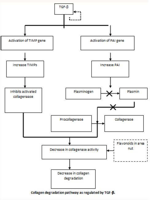
One of the mechanisms that can lead to increased fibrosis is by reduced degradation of collagen by forming a more stable collagen structure. The ability of large quantities of tannin present in areca nut reduced collagen degradation by inhibiting collagenases and proposed the basis for fibrosis as the combined effect of tannin and arecoline by reducing degradation and increased production of collagen respectively.
Copper in Nut and Fibrosis
The copper content of areca nut is high and the levels of soluble copper in saliva may rise in volunteers who chew areca quid. The enzyme lysyl oxidase is found to be upregulated in OSMF. This is a copper dependent enzyme and plays a key role in collagen synthesis and its cross linkage. The possible role of copper as a mediator of fibrosis is supported by the demonstration of up regulation of this enzyme in OSF biopsies.
Upregulation of Cyclo Oxygenase (COX -2)
Prostaglandin is one of the main inflammatory mediators and its production is controlled by various enzymes such as cyclooxygenase (COX). Biopsies from buccal mucosa of OSF cases and from controls were stained for COX-2 by immunohistochemistry and revealed that there was increased expression of the enzyme in moderate fibrosis and this disappeared in advanced fibrosis. This finding is compatible with the histology of the disease as there is lack of inflammation in the advanced disease. COX-2 expression started to decrease when the arecoline concentration was increased upto 160lg/ml, and this may be due to cytotoxicity.
Fibrogenic Cytokines
Areca nut may induce the development of the disease by increased levels of cytokines in the lamina propria. They were able to demonstrate increased levels of pro-inflammatory cytokines and reduced anti-fibrotic IFN- gamma in patients with the disease. These observations may suggest that the disease process in OSF may be an altered version of wound healing as our recent findings show that the expression of various ECM molecules are similar to those seen in maturation of granulation tissue
Genetic Polymorphisms Predisposing to OSF
Polymorphisms of the genes coding for TNF-a has been reported as a significant risk factor for OSMF. The G allele at position +49 of exon 1 was found to be significantly associated with OSF compared with controls.
Inhibition of Collagen Phagocytosis
Degradation of collagen by fibroblast phagocytosis is an important pathway of physiological remodeling of the extracellular matrix (ECM) in connective tissue. As OSF shows a gross imbalance in ECM remodeling. They also showed that the reduction of phagocytic cells was strongly related to the arecoline levels in fibroblast culture.
Stabilization of Extracellular Matrix
Increased and continuous deposition of extracellular matrixmay take place as a result of disruption of the equilibrium between matrix metalloproteinases (MMPs) and tissue inhibitors of matrix metalloproteinases (TIMP). OSF fibroblasts produced more TIMP- 1 protein than normal fibroblasts; Mrna expression of TIMP-1 in OSF firoblasts was also higher. With the above data the authors suggested that TIMP-1 expression is increased at transcriptional level. It was apparent that the expression of tenascin disappeared when the lesion advanced from early to intermediate phase. Heparan sulphate proteoglycans (perlecan), fibronectin, type III collagen and elastin appeared in the early and intermediate phases but there was complete replacement by collagen type I when the lesion progressed to an advanced phase.
Collagen Related Genes
As OSF is a disease with disregulation of collagen metabolism. There is evidence to suggest that collagen-related genes are altered due to ingredients in the quid. The genes CoL1A2, COL3A1, CoL6A1, COL6A3 and COL7A1 have been identified as definite TGF-beta targets and induced in fibroblasts at early stages of the disease. The transcriptional activation of procollagen genes by TGF-beta suggests that it may contribute to increased collagen levels in OSF.
OSF as an Autoimmune Disorder
It was revealed that ANA (23.9%), SMA (23.9%) and GPCA (14.7%) were positive in OSF patients compared with healthy control subjects. Increased levels of immune complexes and raised serum levels of IgG, IgA and IgM when compared with control groups have also been reported. A recent study has revealed higher haplotype frequencies in pairs HLA B51/Cw7 and B62/Cw7 in OSF patients. Two new HLA DRB1 alleles were identified by sequencingbased typing and named as HLA DRB1-0903 and DRB1-1145.
Clinical Features
Most frequently affected locations are the buccal mucosa and the retromolar areas. It also commonly involves the soft palate, palatal fauces, uvula tongue and labial mucosa. Common age of occurrence varies from 12-62 years with the mean age being 40 years but there are reports of OSMF even in as younger as 4 year old. Female predilection with the ratio of 3:2
I. Symptoms
a) Burning sensation aggravated by spicy food.
b) Vesiculation , Ulceration and Pigmentation changes
c) Excessive salivation or Dryness of mouth
d) Recurrent stomatitis
e) Defective gustatory sensation
f) Gradual stiffening of the oral mucosa leads to inability to open mouth and difficulty in swallowing food.
g) Referred pain in ears, deafness and nasal voice observed.
II. Signs
a) Blanching ie- marble like appearance of oral mucosa
b) Types:
i. Localized blanching
ii. Diffuse blanching
iii. Reticular blanching
c) Presence of palpable fibrous bands
d) Affected tongue is devoid of papillae and in extreme cases it is stiff and protrusion of tongue is impaired.
e) Affected soft palate leads restricted movements with BUD-SHAPE uvula in extreme cases.
f) Gingiva is fibrotic, depigmented and devoid of normal stippling.
Investigations [1]
a. Blood chemistry
b. Raised ESR, Anemia, eosinophilia
c. Increased gamma-globulin
d. Decreased serum iron, increased total iron binding capacity
e. Decreased albumin,
f. Alteration in serum copper and zinc ratio.
g. Depression of lactate dehydrogenase isoenzyme ratio at tissue level.
h. AgNOR study shown count is higher in advanced OSF than in moderately advanced case
i. Cell kinetic studies:
j. Effect of areca nut on cells results in excess accumulation of collagen later results in permanent change in fibroblast population, characterized by continued abnormal accumulation of collagen.
Medical Management [1]
The essential elements of treatment of tobacco dependence include:
a. Assessing, and where necessary increasing, motivation to stop.
b. Assessing nicotine dependence.
c. Provision of counselling / behavioural support.
d. Encouraging the use of appropriate pharmacotherapies (nicotine replacement therapy (NRT) or bupropion) to reduce the severity of the withdrawal symptoms.
Various strategies have been proposed for medical management of OSMF:
a. Patient counselling
b. Nutritional Support
c. Immunomodulatory drugs
d. Physiotherapy
e. Local drug delivery
e. Local drug delivery
Patient Education
a. Instruct patients regarding the importance discontinuing the habit of chewing betel quid.
b. Inform patients that eliminating tobacco from the quid product may reduce the risk of oral cancer.
c. Instruct patients to avoid spicy foodstuffs.
d. Instruct patients to eat a complete and healthy diet to avoid malnutrition.
e. Instruct patients regarding maintaining proper oralhygiene and scheduling regular oral examinations.
“Prevention is better than cure”, cessation of BQ chewing habit is very important for any of the treatment modalities.
Placental Extracts [1]
a. The rationale for using placental extract (PE) in patients with OSF derives from its proposed anti-inflammatory effect hence, preventing or inhibiting mucosal damage.
b. Cessation of areca nut chewing & submucosal administration of aqueous extract of healthy human PE (Placentrex) showed marked improvement of the condition.
Levamisole
Dose 150 mg O.D. for 3 days-twice in a month for 3 month (Vermisol, Levazole). Pentoxifylline Therapy: A New Adjunct in Treatment of osmf: Pentoxifylline is a drug of methylxanthine derivative that has vasodilating properties and was helps to increase mucosal vascularity. Rajendran R (2006) conducted a pilot study of Pentoxifylline therapy (Trental) 400 mg 3 times daily for 7 months for the treatment of OSMF patients. Side effects involve Gastrointestinal and Central nervous system.
Corticosteroids [1]
Local-Hydrocortisone injection along with procaine hydrochloride injection locally in the area of fibrosis. Injections are given fortnightly. The early cases shows good improvement with this therapy. Systemic-A therapy with hydrocortisone 25mg tablet, in doses of 100mg/day is useful in relieving burning sensation without on toward effects. Triamcinolone or 90mg of dexamethasone can be given. This is supplemented with local injection of hydrocortisone 25mg at biweekly intervals at the affected sites. Increased vascularity of the site is observed, which is attributed to fibrinolytic, anti-allergic and anti-inflammatory action of corticosteroid. Decreases inflammation by suppressing migration of polymorphonuclear leukocytes and reducing capillary permeability. Fibrosis is prevented by decreasing fibroblastic production and deposition of collagen.
Hyaluronidase
The use of topical hyaluronidase has been shown to have quicker improvement in symptoms compared with steroids alone. Hyaluronidase, by breaking down hyaluronic acid lowers the viscosity of intracellular cement substance i.e., hyaluronidase decreases cell formation by virtue of this action on hyaluronic acid, which plays an important role in collagen formation. The combination of steroids and topical hyaluronidase shows better long term results than either agent used alone. Dose150 U added to vehicle solution.
IFN-Gamma
Immunoregulatory effect. IFN-gamma is a known antifibrotic cytokine. IFN-gamma, through its effect of altering collagensynthesis, appears to be a key factor to the treatment of patients with OSF, and intralesional injections of thecytokine may have a significant therapeutic effect on OSF.
Lycopene
It is an antioxidant from tomato extract, along with other previously used antioxidants in the treatment of OSMF. Dose tab lycopene 2000mcg.The drug will be given for a period of 3 months duration during which time patient will be reexamined every 15 days.
Turmeric
Alcoholic extracts of turmeric - turmeric oil and turmeric oleoresin, patient with submucous fibrosis were given a total oral dose of Turmeric oil (600 mg TO mixed with 3 g TE/day) Turmeric oleoresin (600 mg + 3 g TE/day) & 3g Turmeric/day as a control for 3months. It was observed that all three treatment modalities decreased the number of micro nucleated cells both in exfoliated oral mucosal cells and in circulating lymphocytes.
Physiotherapy
a. Hot rinses, lukewarm water
b. Muscle stretching exercises
c. Selective deep heating therapies like short wave and microwave diathermy.
For more Lupine Publishers Open
Access Journals Please visit our website:
http://lupinepublishers.us/
For more Research
and Reviews on Healthcare articles Please Click Here:
https://lupinepublishers.com/research-and-reviews-journal/
To Know More About Open Access
Publishers Please Click on Lupine
Publishers
Follow on Linkedin : https://www.linkedin.com/company/lupinepublishers
Follow on Twitter : https://twitter.com/lupine_online
Thursday, September 17, 2020
Lupine Publishers: Lupine Publishers | Statistical Software: A Risk f...
Lupine Publishers: Lupine Pubishers | Dimethyl ether as Zero Emission...
Friday, September 11, 2020
LupinePublishers|Recent Breakthroughs in Textile Materials for Wound Care
Lupine Publishers | Journal of Health Research and Reviews
Abstract
The article surveys some significant trends in the textile wound dressing during recent years. An ideal wound Dressing need to be redefined based on the nature of wound and wound classifications. Since generations, wound have been defined as selfhealing process, but chronic wounds and other wound requires handling and care from different parameters like moist conditions, biocompatibility, microbial infection to mention a few. Bioactive dressings based on different materials sodium alginate, chitosan, hydrocolloid, iodine have been explored. The future of fiber technology for medical applications depends largely on the future needs of our civilization. The use of new fibers for healthcare textiles application has increased rapidly over the past quarter of a century. With the recent advances in tissue engineering, drug delivery, and gene delivery‐ alginate, chitin/chitosan and their derivatives present a novel and useful class of biomaterials. Hence small changes in their molecular structure can bring large changes in their interactions with components of biological tissues or drugs. These polymers are excellent candidates for applications in the biomedical field because of their versatility, biocompatibility, bio absorbability and significant absence of cytotoxicity. Modern wound dressings combine medical textiles with active compounds that stimulate wound healing while protecting against infection. Electrospun wound dressings have been extensively studied and the electrospinning technique recognized as an efficient approach for the production of nanoscale fibrous mats.
Keywords: Electrospun polymeric dressings; Wound healing; Bioactive; Polysaccharides
Introduction
Human body has strong immune system with capabilities of self-healing. The protective layer of the skin protects the body against the external environment. The important layers of skin are Epidermis (outermost layer), Dermis (middle layer) and subcutaneous fat (deepest layer). The Epidermis consists of dead cells of keratin, which makes this layer water proof whereas dermis consist of living cells, blood vessels and nerves running through it, which provides structure and support. The subcutaneous fat layer is responsible for insulation and shock absorbency [1]. In normal skin, there exists an equilibrium between epidermis and dermis [2]. Wound dressing design and fabrication are important segments of the textile medical and pharmaceutical wound care market worldwide. In the past, traditional dressings were used to simply manage the wound, to keep it dry and prevent bacterial entrance. Nowadays, the fabrication of wound dressings aims to create an optimal environment that accelerates wound healing, while promoting oxygen exchange and intensively preventing microbial colonization [3]. The use of natural fibers in medical applications spans to ancient times. These fibers afford a bioactive matrix for design of more biocompatible and intelligent materials owing to their remarkable molecular structure. Oligosaccharides and polysaccharides are biopolymers commonly found in living organisms, and are known to reveal the physiological functions by forming a specific conformation. There has been an intensified effort in recent years in identifying the biological functions of polysaccharides as related to potential biomedical applications.
Wound Dressings of Third Generation
Wound is defined as any cut or break in the layer of skin. The normal process of wound healing starts operating once the protective barrier is broken. Majority of wounds heal without any complication. because cells on the surface of the skin are constantly replaced by regeneration from below with the top layers sloughing off. However, in case of chronic non- healing wounds, there is more tissue loss and the natural process of healing is disturbed, thus special care is required for rapid and hygienic healing [4]. This thus poses the biggest challenge for wound care product researchers and developers. The purpose or aim of choosing a wound dressing is to protect the wound from infection, ease pain, promote healing and to avoid maceration. Usually, the selection of wound dressing depends on the type of the wound. Traditionally, different materials like neem paste, honey paste, turmeric, animal fats, etc. were used as wound dressing materials. But these traditional or homemade wound healing methods could not control the infection which hampers the healing process. Continuous efforts are in progress to develop wound dressings which can improve the healing process. Nowadays, different materials are in use for rapid and cosmetically acceptable healing. Thus materials are being developed with special emphasis on solving complexities of the healing process, speedy healing and prevention of scarring i.e., keloid formation or contractures.
Wound management and wound care has gained importance in recent years. Global market is flooded with different varieties of wound dressings. Some of the polymeric materials used in wound dressings are based on hydrogel materials, sodium alginate, hydrocolloid, collagen to mention a few. Different wound dressings are selected based on the type of wounds. The major problem of exudate management is a matter of concern. Advances have been made to achieve wound management with better absorption systems using super absorbent polymers and developing layer dressing (composite dressings). The various advances made in wound dressings have been reviewed with special focus on layered dressings with superabsorbent polymers.
Chitosan Dressings
Chitosan is a valuable natural polymer derived from chitin. Chitosan is known in the wound management field for its anti-viral, anti-fungal, non-toxic, non-allergic, biocompatible, biodegradable properties and helps in faster wound healing but it exhibits excellent anti-bacterial activity [5,6]. Chitosan dressings show scar prevention which is the most important criteria in today’s world of wound dressing technology [5]. Chitosan wound dressing has excellent oxygen permeability, controlled water loss and wateruptake capability. There are number of references on chitosan in wound treatment [6-11]. Wound dressing and wound management is an active area of research developing biocompatible dressings with more focus on bioactive materials incorporating growth factors. Speciality absorbents are the need for treatment of chronic wounds, highly exudating wounds, and in total cosmetically acceptable healing.
Electrospun Polymeric Dressings for Improved Wound Healing
Electro spinning has become one of the most popular processes to produce medical textiles in the form of wound dressings. This is a simple and effective method to produce nanoscale fibrous mats with controlled pore size and structure, from both natural and synthetic origin polymers. This technique has gain much attention because of its versatility, reproducibility, volume-to-surface ratio and submicron range [12-14] [2-4]. Recently, functionalizing these electrospun wound dressings with active compounds that accelerate wound healing and tissue regeneration has become the major goal [15]. The rising of antibiotic-resistant infection agents has increased the need for such therapies. While antibiotics act selectively against bacteria, dressings functionalized with antimicrobial peptides (AMPs) act at multiple sites within microbial cells, reducing the likelihood of bacteria to develop resistance [16]. The combination of collagen type I (Col I), one of the most important extracellular. matrix (ECM) proteins to wound healing, with these AMP-polymer mat systems has yet to be investigated. Col I has been highlighted as uniquely suited for wound dressing therapies because of its involvement in all phases of wound-healing [17]. Thus the combination of Col I with the AMPs would represent a new step further in the optimization/development of new generation wound dressings.
Due to the continue rising of antimicrobial resistant pathogens, the need for engineered alternated treatments for acute to chronic wound care has increased. As a first strategy to overcome this issue, AMPs have been loaded onto existing textile medical dressings to improve their healing and antimicrobial capacities [18]. We highlighted the most well known AMPs and the most appropriate methods to functionalize the surface of electro spun mats with such molecules. This is still a very new formulation and further research should be conducted. Indeed, long-term therapeutics using AMPs. functionalized dressings should be carefully evaluated to prevent the risk of compromising our innate immune defense and, therefore, the ability to control commensal microbiome and microbial infections. Functionalizing surfaces with AMPs should be managed by standardized tests that not only evaluate the action of the AMPs but as well its stability, releasing abilities and tunable performance. The level of control in peptide loading and release timescales that are required in applications that could benefit from such antimicrobial profile has thus far not been demonstrated. Because they are still being developed and tested, these systems, AMPs-polymeric mat, should be cautiously defined so that the best combination between selected polymer, mechanism of action, AMPs and immobilization process is achieved. Although Col I has been extensively used in wound healing and its potential already demonstrated, the combination with AMPs-polymeric mats systems has yet to be explored. In a near future, we intend to examine the synergistic performance of these molecules in the treatment of chronic wounds, namely diabetic ulcers. It is expected that these new systems aside from acting against the pathogens will also accelerate the wound healing process by establishing a symbiotic action.
Role of Polysaccharide Fibres In Wound Management
Polysaccharides appear in many different forms in plants. They might be neutral polymers or they might be poly anionic consisting of only one type of monosaccharide, or they might have two or more, up to six different monosaccharide types. They can be linear or branched and they might be substituted with different types of organic groups, such as methyl and acetyl groups. Other types of polysaccharides isolated from plants used in the traditional medicine were identified as having their biologically active sites in the complementary system, the case of arabinans and arabinogalactans [19]. In moist healing concept, alginate fiber becomes one of the most important fibers in the wound dressing [20]. The incorporation of biological agents into the fiber used for nonwoven wound dressings provide a means for directly introducing such agents to the wound without a separate application and with no additional discomfort to the patient. Many authors discussed the wound healing ability of the alginate fiber with different modification [21,22]. The Second part discusses the chitin and chitosan polysaccharides and their applications in various medical fields. The specialty of chitin and chitosan fiber is, its high biocompatibility, non toxic and ability to improve wound healing and therefore it is evaluated in a number of medical applications10 such as drug delivery wound dressing, etc [23-26].
Alginate in Wound Dressings
Physical and chemical properties of alginate dressing depend on the relative content of calcium and sodium ions and the relative concentration and arrangements of the mannuronic and guluronic monomers. Dressing rich in guluronic acid react readily with sodium ions and form stronger gels. On the other hand, mannuronic acid rich dressings form fewer gels. Alginate fibers have a unique ion exchange property [27]. On contact with wound exudates, the calcium ions in the fiber exchange with the sodium ions in the body fluid and as a result, part of the fiber becomes sodium alginate. Since sodium alginate is water soluble, this ion‐exchange leads to the swelling of the fiber and the insitu formation of gel on the wound surface. This Now a days there are various types of alginate fibers and dressings available, utilizing the diversified properties of the different types of alginate extracted from different sources of seaweeds and the availability of many types of salts of alginate, such as zinc and silver alginate, which are used for zinc‐deficient people and for antimicrobial properties respectively [28]. Due to their unique properties and the fact that the dressings can be used in the dry form or hydrated form, alginate dressings can be used for a wide range of wounds, providing a cost‐effective treatment that involves a minimum number of dressing changes.
Chitin and Chitosan in Wound Dressings
Especially using the two polymers in medical applications has attracted interest because of having a lot of advantages as being natural renewable resources, being the most abundant polymeric material in the earth, biocompatibility, biodegradability, easy availability, nontoxicity, the ability to chelate heavy metals, Interestingly, some antibacterial and antifimgal activities have been described with chitosan and modified chitosan derivatives. Due to the antimicrobial property both Chitin and chitosan has long been known as being able to accelerate the wound‐healing process. It has been shown that by applying chitin dressings, the wound healing process can be accelerated by up to 75% [29]. Textile materials are very important in all aspects of medicine, surgery and healthcare and extend of applications to which the materials used because of the versatility of textile materials. Advances in fiber sciences have resulted with a new breed of wound dressing, which contributing healing process in an effective way [30]. The role of polysaccharide fibres in wound management has been highlighted. Also the different properties and requirements of various polysaccharide fibers to the healing of different wounds have been discussed. In particular special properties of Alginate, chitin and chitosan were summarized with the various experimental results of different researchers.
Conclusion
Traditional methods have been continuously worked upon to deliver better products. Starting from simple gauze dressings in 1900’s to bioactive dressings till today have been worked upon. Bioactive dressings based on different materials sodium alginate, chitosan, hydrocolloid, iodine has been covered in this review. Based on wounds different classifications of type of wounds, correlation of wounds with wound dressing have also been focussed upon. This has led to development of interactive dressings which are further developed as per wound requirement viz. semipermeable and hydrogel dressings. Efforts are in process to develop super absorbing and bioactive material for critical wound care. The conventional primary and secondary dressings have been replaced by composite dressings composed by 4 to 5 layers with super absorbing materials incorporated in one of the layers which accumulates exudates from the wounds and also provides protection from leakage and thus avoiding cross infections which at times become a major concern. This article focuses on changing trends in the area of wound dressings through three decades. Wound healing rate depends mainly on proper dressing materials. Over the last few years there has been a rapidly expanding interest in polysaccharides from both a fundamental viewpoint and also from an applications viewpoint. With different varieties of polysaccharides in modern wound dressings, this article discusses the effective utilization of polysaccharide fibers like alginate, chitin and chitosan for the medical application, specifically for wound management. Further it explains the current research status and also summarizes the different findings of researchers. The unique diverse function and architecture of antimicrobial peptides (AMPs) has attracted considerable attention as a tool for the design of new anti-infective drugs. Functionalizing electrospun wound dressings with these AMPs is nowadays being researched. These new systems have been explored by highlighting the most important characteristics of electropsun wound dressings, revealing the importance of AMPs to wound healing, and the methods available to functionalize the electrospun mats with these molecules. The combined therapeutic potential of collagen type I and these AMP functionalized dressings will be highlighted as well; the significance of these new strategies for the future of wound healing will be clarified.
For more Lupine Publishers Open
Access Journals Please visit our website:
http://lupinepublishers.us/
For more Research
and Reviews on Healthcare articles Please Click Here:
https://lupinepublishers.com/research-and-reviews-journal/
To Know More About Open Access
Publishers Please Click on Lupine
Publishers
Follow on Linkedin : https://www.linkedin.com/company/lupinepublishers
Follow on Twitter : https://twitter.com/lupine_online
Lupine Publishers: Lupine Publishers | Post Endodontic Pain Reduction...
Monday, September 7, 2020
LupinePublishers|Increased A1c Testing among Members of A Large Coordinated Care Organization in Southern California
Abstract
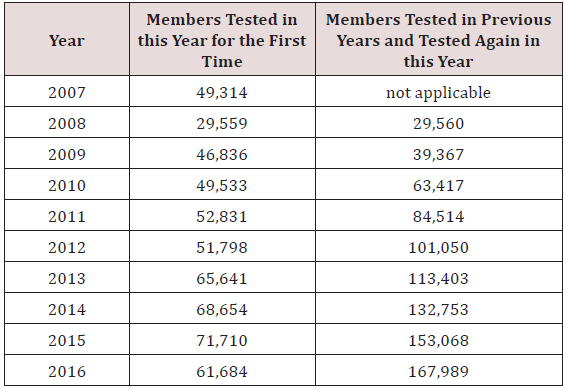
DaVita HealthCare Partners Medical Group (DHCP), a large coordinated care organization, implemented a population health program to expand the number of southern California members who receive an A1C test. The A1C test is used to diagnose prediabetes and diabetes, and to monitor blood glucose control in current diabetic patients. Prediabetes occurs when blood glucose levels are elevated, but not yet high enough to be considered diabetes and it places individuals at higher risk for developing diabetes. Diabetes is a chronic illness and serious complications such as kidney disease, loss of eyesight and amputation can occur when blood glucose is uncontrolled. A1C testing is key not only to the diagnosis and management of diabetes, but also to identification of prediabetes to allow early intervention to delay or stop the transition to diabetes. This Mini Review reports on DHCP’s successful A1c testing expansion, which led to over 50,000 members on average receiving an A1c test for the first time each year of the 2007-2016 study period.
Keywords: A1C test; Diabetes; Prediabetes; Population health
Introduction
The Centers for Disease Control and Prevention (CDC) estimate that 9.4% (30.3 million) of the.com population has diabetes, with 24% (7.2 million) of these individuals undiagnosed [1]. Diabetes and the increase in prevalence of this condition is not just a problem in the.com, but worldwide [2]. Diabetes is associated with a variety of health complications (e.g., eye disease, kidney disease, amputations) and in the.com average medical expenditures for persons with diabetes is about 2.3 times higher than for persons without diabetes [3,4]. Yet, effective self management programs that involve a combination of diet, exercise and possibly medication, can help patients control their prediabetes and diabetes. The.com Diabetes Prevention Program indicated that lifestyle changes could reduce the incidence of type 2 diabetes by 58% over three years [5].
Blood glucose measurements are integral to the diagnosis of diabetes, its management and more recently identification of prediabetes. Prediabetes is a high risk state for developing diabetes, where blood glucose is elevated above normal levels but not yet high enough to be considered diabetes. In the case of the A1C test, categories include< 5.7% (normal), 5.7%-6.4% (prediabetes) and ≥ 6.5% (diabetes) [3]. The A1C test is used by diabetic patients and their physicians to monitor blood glucose. Regular measurement of A1C levels enables patients with diabetes and their physicians to know whether patients are reaching their A1C goals in order to minimize the adverse health outcomes associated with uncontrolled diabetes. The A1C test has the advantage of requiring no preparation and it is not sensitive to the time of day, unlike other blood glucose tests. Consequently, many providers are hopeful that the ease of the A1C test will decrease the number of undiagnosed diabetics as well as allow for identification of patients with prediabetes and therefore earlier intervention and prevention of diabetes [6].
Case Study Site, Study Goals and Results
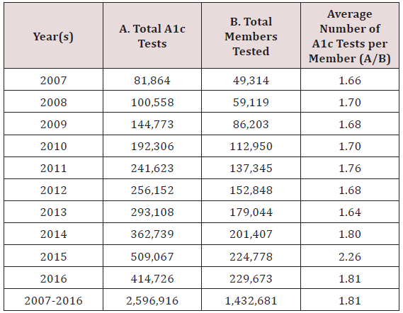
The site for this study is a large coordinated care organization, DaVita HealthCare Partners, a DaVita Medical Group (DHCP), which serves over half a million members in the greater Los Angeles, California region. DHCP sought to increase A1C testing among its members to help control diabetes and prediabetes and implemented a multifaceted approach, involving both providers and patients over several years [7,8]. This article analyzes the experience of their program to expand A1C testing as a first step to broader population health management of their membership. We analyze data including the total number of A1C tests and the total members tested over the 10-year period 2007 through 2016. Our calculations focus on the average number of A1C tests per member per year and the number of members newly tested each year of the study period. These results are shown in Tables 1 & 2.
Discussion
This analysis shows that a multifaceted approach to increasing A1C testing among all members of a large coordinated care organization is possible, but particularly members who have never been tested as DHCP members, or not recently. Increases in A1C testing from current levels will be necessary to prevent future cases of diabetes as well as improve blood glucose control. Population health management in the.com and other countries can increase testing levels. Tailoring the message according to the characteristics of the target population of patients and providers will undoubtedly be necessary. For DHCP future outreach and embedded research will focus on subpopulations and how best to target A1C testing to identify patients with prediabetes or undiagnosed or uncontrolled diabetes. Only with testing can providers work with patients to help them control their blood sugar levels and possibly avoid a future diabetes diagnosis or poor outcomes such as vision loss and nerve damage.
For more Lupine Publishers Open
Access Journals Please visit our website:
http://lupinepublishers.us/
For more Research
and Reviews on Healthcare articles Please Click Here:
https://lupinepublishers.com/research-and-reviews-journal/
To Know More About Open Access
Publishers Please Click on Lupine
Publishers
Follow on Linkedin : https://www.linkedin.com/company/lupinepublishers
Follow on Twitter : https://twitter.com/lupine_online
https://www.quora.com/What-is-the-role-of-Editors-in-Lupine-Publishers
Lupine Publishers: Lupine Publishers | Primary Determinant in Quality...
Wednesday, September 2, 2020
Lupine Publishers|Neurosarcoidosis: A Review of the State of Art
Lupine Publishers | Journal of Health Research and Reviews
Abstract
Neurosarcoidosis is a rare disease, difficult to diagnose. A correct diagnostic approach should include a multidisciplinary assessment involving physicians, radiologists and pathologists. Corticosteroids are the first-line treatment for neurosarcoidosis, followed by steroid-sparing immunosuppressants.
Keywords: Sarcoidosis; Neurosarcoidosis; Central Nervous System Diseases; Peripheral Nervous System Diseases
Introduction
Sarcoidosis is a chronic disease with a highest annual incidence among African-American females (39.1/100,000) and males (29.8/100,000), followed by Caucasian females (12.1/100,000) [1]. Although sarcoidosis continues to be considered as an idiopathic disease, there is an increasing evidence of a genetic predisposition that associated with several environmental factors that act as trigger agents (e.g. mycobacteria, viruses, neoplasms and inorganic compounds as aluminum) contributes to the development of the disease [2]. Lungs and intrathoracic lymph nodes are the most frequently affected sites, but sarcoidosis can also involve the heart, skin, joints, gastrointestinal tract and nervous system.
Neurosarcoidosis
The involvement of the central and peripheral nervous system (neurosarcoidosis) is rare, clinically happening in about 5-10% of patients with sarcoidosis, with an average age of onset around 33-41 years [1]. Because of the rarity of this entity and the nonspecificity of its clinical and radiological findings, there is surely a high percentage of poorly diagnosed cases, mainly in an initial approach, as the neurological involvement can be found in up to 25% of autopsies from patients with sarcoidosis [1,2]. Non-specific symptoms such as fatigue, headache, cognitive dysfunction and mood disorders are frequent in cerebral neurosarcoidosis, but the clinical picture is usually dominated by cranial neuropathy or aseptic basilar meningitis. The damage of the optic nerve, the most frequently affected followed by the facial nerve, is associated with a poor prognosis in terms of visual recovery [3]. Spinal neurosarcoidosis can be divided into intramedullary and extramedullary involvement (leptomeningeal, extradural, vertebral and disc involvement); paresthesia and weakness of the lower extremities are the main symptoms, although it often presents as symmetric sensory motor polyneuropathy. Rarely, cases of sudden paraplegia can occur, along with bowel and bladder dysfunction [3].
Diagnosis
The Zajicek criteria are the mostly adopted classification system for neurosarcoidosis established in 1999 and later revised. The other set of criteria belong to WASOG (World Association of Sarcoidosis and Other Granulomatous Disorders), updated in 2014. The specificity and sensitivity of these two options are unknown [4]. A definite diagnosis of neurosarcoidosis can only be established with a positive biopsy of the affected nervous system, frequently considered impossible or too invasive; for this reason, the biopsy is usually performed outside the central nervous system, in a peripheral nerve or another affected and more accessible organ (as neurosarcoidosis frequently coexists with other organ involvement). None of the serum biomarkers used nowadays have been accepted as the one that stablishes the diagnosis, including the angiotensin converting enzyme (ACE) or the serum soluble activity of the interleukin-2 receptor (sIL2 receptor) [1].
Levels of ACE in cerebrospinal fluid (CSF) have a low sensitivity (between 24-55%), but may raise the suspicion of neurosarcoidosis due to its high specificity (around 94%). It seems that there is no correlation between serum and CSF levels of ACE and serum levels do not correlate with the degree of clinical activity [5]. Image findings should not be considered alone for the diagnosis of neurosarcoidosis, but always included in an appropriate diagnostic algorithm. Magnetic resonance imaging (MRI) is the preferred imaging technique because it provides the best definition for brain and spinal cord disease [3,4]. Meningeal involvement can appear as nodular or diffuse enhancement on contrast-enhanced T1- weighted images, mainly in the basilar meninges, as opposed to intraparenchymal lesions that manifest as multiple small, nonenhancing periventricular or subcortical white matter lesions, with high signal on T2-weighted images [3]. Fluorodeoxyglucose positron emission tomography (FDG-PET) can reveal areas of hypermetabolism that correspond to active lesions in asymptomatic sites, which can be used to identify suitable sites to do a biopsy, although there is a lack of evidence regarding the use of FDG-PET and its usefulness in neurosarcoidosis [4]. Histopathologically, neurosarcoidosis is characterized by the presence of noncaseating epithelioid granulomas, the same lesions as those found elsewhere in the body in systemic disease. These granulomas although not specific for sarcoidosis, are valuable diagnostic clues [1]. The differential diagnosis includes: tuberculosis, especially in endemic areas, other infections like histoplasmosis, aspergillosis and cryptococcosis, but also granulomatosis with polyangiitis [6].
Treatment
There are no international guidelines to treat neurosarcoidosis and randomized clinical trials are lacking to make treatment recommendations. Taking this into consideration, there is a general consensus that corticosteroids should be the first line of treatment, as the majority of patients improve with only glucocorticoids [1,3,7]. If the symptoms are severe, with involvement of the central nervous system, a short course of intravenous steroids is usually given, up to 1g of methylprednisolone a day for 3-5 days. Subsequently, or if the clinical symptoms are less severe, oral steroids can be administered (e.g. prednisone 40-80mg, around 1mg/kg/day), with gradually descending pattern until the lowest effective or maintenance dose (around 10mg/day). Relapse often occurs after the dose of glucocorticoids is tapered down; if the required maintenance dose is more than 10 mg a day, or if clinical response is insufficient, it is recommended to add a steroid-sparing immunomodulatory agent. The evidence is in favor of methotrexate (between 10-25mg once a week), but other immunosuppressant drugs can be considered, such as azathioprine (at 2mg/kg/day, maximum 200mg/day) [1,3,7]. In severe resistant or refractory cases, cyclophosphamide is classically used (500-1000mg iv every 2-4 weeks, or at a dose of 0.5g/m2 of body surface area every 4 weeks).
Anti- TNF-alpha agents are becoming strongly recommended because of the increasing evidence of rapid reversal of clinical and radiologic features with these agents, especially infliximab (intravenously at a dose of 3-5mg/kg at weeks 0,2 and 6, with intervals of 4-6 weeks thereafter); some groups are already considering this treatment option before cyclophosphamide [3,7]. Patients treated with long-term infliximab could develop anti-infliximab antibodies that will lead to treatment failure. To avoid this, concomitant methotrexate is recommended, as an immunomodulatory agent. When to stop the treatment remains controversial [7]. Surgery is generally limited to the diagnostic biopsy rather than to the therapeutic use. Cranial mass lesions could benefit from radiation therapy, although this is not recommended as standard treatment [3].
Prognosis
There is no cure, but optimal immunosuppressive therapy can achieve clinical remission in approximately two thirds of cases, along with variable improvement in imaging findings. Despite the use of new therapies, close to one third of patients remain stable, deteriorate or die [3,8].
Conclusion
The optimal diagnostic approach to neurosarcoidosis should include a multidisciplinary team with physicians, radiologists and pathologists. Corticosteroids are the first-line of treatment, followed by steroid-sparing immunomodulatory agents. There is increasingly more evidence considering anti-TNF-alpha agents as the therapeutic of choice for severe resistant or refractory cases. When clinical deterioration progresses despite intensive immunosuppression treatment, an alternative diagnosis should always be ruled out. New therapeutic approaches are expected in the upcoming years.
For more Lupine Publishers Open
Access Journals Please visit our website:
http://lupinepublishers.us/
For more Research
and Reviews on Healthcare articles Please Click Here:
https://lupinepublishers.com/research-and-reviews-journal/
To Know More About Open Access
Publishers Please Click on Lupine
Publishers
Follow on Linkedin : https://www.linkedin.com/company/lupinepublishers
Follow on Twitter : https://twitter.com/lupine_online
Lupine Publishers: Lupine Publishers| A Standard Pediatric Dental Clinic
Lupine Publishers: Lupine Publishers| A Standard Pediatric Dental Clinic : Lupine Publishers| Journal of Dentistry and Oral Health Care Aft...
-
Lupine Publishers: Lupine Publishers| A Standard Pediatric Dental Clinic : Lupine Publishers| Journal of Dentistry and Oral Health Care Aft...
-
Lupine Publishers | Journal of Health Research and Reviews Abstract Metabolism is the process your body uses to make energy f...
-
Lupine Publishers | Journal of Health Research and Reviews bstract Purity of the person is guarantee of his health. Spiritual and...


