Showing posts with label Medical and Health Sciences. Show all posts

Friday, April 22, 2022

Lupine Publishers|An Innovative Method for Endovascular Stabilization of Vulnerable Plaque in Coronary Arteries: An Opinion


  Lupine Publishers | Journal of Health Research and Reviews


Short Communication

Despite all of the available diagnostic and treatment modalities atherosclerosis remains one of the most common healthcare problems worldwide with an estimated annual mortality rate of approximately 17,5 million cases [1]. In most cases acute coronary syndrome (ACS) appears to be linked to atherosclerotic lesion associated thrombosis of a vessel [2]. In the situation, when patients come to the interventional cardiology unit with a confirmed diagnosis of ACS: myocardial infarction, a so-called culprit lesion can be identified and treated according to international guidelines. However, alongside the lesions, that are obviously causing an impairment of a blood perfusion in a certain segment of the myocardium or have already destabilized causing acute thrombosis, some other form of entity can frequently be seen. These are called non – culprit lesions. Even though their appearance in an orifice of a vessel is regarded by most surgeons as a “bad omen”, the international community is still puzzled and unsure, if these lesions are to be treated. And even if they are, the specialists cannot yet be sure, what type of an intervention is preferable. This problem arises from several premises, including the following:

I. The non – culprit vulnerable plaque destabilization is a complex phenomenon. Not only the mechanical properties and structural integrity of the plaque define the further events, but the mechanical forces [3-5], that affect the plaque and are being transduces by blood flow, a non-Newtonian fluid, whose properties might be affected in a large variety of conditions. Apparently, the hemostasis must be accessed prior to drawing the right decision in patient’s follow – up tactics. But exact parameters are yet to be determined [6].

II. The non – culprit vulnerable plaque destabilization does not always cause an ACS [7]. Some specialists imply that assessment of blood properties and structural characteristics of the plaque are not enough. Apparently, the state of a myocardium must also be assessed.

III. Not all vulnerable plaques are equal, and the risks of destabilization accompanied by MACE vary depending on the plaque localization [8]. This statement appears to be obvious. Most surgeons would have guessed this without any researches: the more approximate position in a coronary artery clearly indicates a higher chance of an unfavorable outcome. However, what might be not so apparent, the different segments of coronary arteries’ do not react to sheer-stress in a uniform manner [9]. This is almost impossible to assess in real clinical practice but is a good thing to bear in mind.

One must put a lot of effort into the diagnostic procedures, in order to understand the whole situation. Simultaneously we do not know what kind of a medication must be used in every situation. A lot of effort has been put recently into finding a preferable drug for stabilization of a vulnerable plaque. Different approaches were used. Considering the trails REVERSAL, SATURN, ASTEROID and most importantly YELLOW, the conventional statins are still a medication of choice for the most patients with vulnerable plaques. Other drugs were introduced recently, including ivabradine [10], grelin [11], canakinumab [12]. Most of these approaches are still in early development, but it is clear now, that the lack of preventive conservative treatment is a risk factor for any manipulation [13], that is to be performed upon a vulnerable plaque.

Some cardiologists imply that more aggressive invasive techniques should be prioritized. We possess only scarce data. PRAMI trial indicates, that stenting any lesion, that is even less than 50% might be beneficial [14]. The CvLPRIP trial has mostly confirmed the findings of PRAMI [15]. Interesting results were obtained by Dai et al [16] they were able to demonstrate, that routine stenting of all lesions leads to lower rates of death, secondary ACS. MACCE were lower: RR (HR) 0.35 [95% CI 0.18 – 0.69]. Additional factors listed above might contribute to drawing a right decision in different situations.

The problem with invasive procedures extends beyond that:

I. The risks of stent - thrombosis are unacceptable in endovascular plaque stabilization. This in turn requires at least using an extremely precise tool, that has only limited contact with endothelial cells, that have not yet succumbed to pathological process. Extensive damage frequently leads to neointimal hyperplasia, neoatherosclerosis and stent thrombosis. Considering all things stated, we suppose, that only truncated stents might be used for vulnerable plaque stabilization.

II. The usage of truncated stents requires a fairly good level of precision, that most devices nowadays are still unable to provide.

III. The risks of bleeding are also high, because a person is forced to take a variety of anticoagulants for a prolonged period after the procedure. The bigger the time interval, the bigger the chances of a major bleeding. Therefore, there is a question to be asked: what if the biodegradable stents, that did not legitimately receive much attention due to the known issues and inability to compete with DES in terms of conventional stenting, can be used in this situation.

IV. The risks of periprocedural myocardial infarction [17] are dependent on structural properties of the plaque. High lipid burden and large lipid core are mostly responsible for this adverse event [18]. This cannot be manipulated by the means of surgical instruments and is a single reason for carefully assessing the situation using different diagnostic techniques.

V. It is economically ineffective to create a separate device for treatment of vulnerable plaques only.

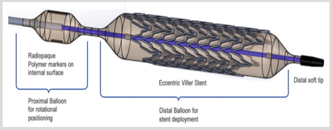
VI. Bearing in mind all the concerns stated, we decided to come up with a new device that can be used in many different fields but is also capable to satisfy all the precautions involved in vulnerable plaque endovascular stabilization. The throughout description of the basics of this over the wire stent delivery systems (SDS) construction is not however the goal of this article. All the information can be found in our patent here: US 20100070014 A1 published in 2010. The project is in early development and we do not encourage a reader to buy it. Therefore, this article is not a commercial, but a call to international specialists, that would probably find this topic interesting to discuss. The basic structure of the SDSs distal shaft is shown below (Figure 1).

Figure 1.

Lupinepublishers-openaccess-Research-Reviews

Such SDS works in following order. Both balloons are connected consecutively to the compressor, but radiopaque label bearing balloon is more compliant and expands in the first place allowing the precise positioning of asymmetrical truncated stent in the orifice of the vessel. By applying sufficient force, the operator can ensure that the stent is in position, meanwhile, by applying additional pressure, can start the expansion of the second stent- bearing balloon. After the implantation is complete the SDS is removed at once. We are currently developing new biodegradable stents and will test the whole system on a swine model in several months.

For more Lupine Publishers Research and Reviews on Healthcare Open Access Journal Articles:
https://lupine-publishers-research.blogspot.com/


Monday, August 23, 2021

Lupine Publishers| Task Acquisition and Motor Learning Require Conscious Efforts by The Patients


  Lupine Publishers | Journal of Health Research and Reviews


Motor learning and skill acquisition is a complex process of learning through the interaction of various dependent variables. One of the most important variables is the conscious effort of the patients to learn a new skill or to refine a learned skill. Social and communication skills of a therapist play an important role in motivating and engaging cognitionof a patient to learn a new motor skill.

Keywords:Motor Learning; New Skill; Practice; Training

Motor learning is a “collection of internal processes associated with practice leading to relatively permanent changes in the skilled behavior.” In other terms, motor learning is when complex processes occurring in the brain, in response to the practice of a particular skill resulting in the change in neuronal network [1]. Motor learning involves three stages such as, cognitive, associative and autonomous [2]. Except for the autonomous stage, cognitive and autonomous stage requires a conscious effort by the patients [3-5]. The Smart Approach (SA) is a systematic way to approach a patient for better rehabilitation outcome. The SA involves the following stages.

a. Stage 1: Reviewing the medical record of the patient.

b. Stage 2: Review the latest scienti􀏐ic literature for the best current practice.

c. Stage 3: The rehabilitation goals must be formulated in consultation with the patients and care takers.

The SA improves patients trust in therapist, which is essential factor in the success of the treatment. SA by a therapist would result in greater acceptance of activity modi􀏐ication and adherence to the regime, lower anxiety level in relation to any con􀏐lict [6-9]. Patients are more likely to open and disclose information if they trust their therapist. [6] [10,11]. In view of the above, it’s imperative for a therapist to gain the con􀏐idence of the patients through a SA. Regular one to one personal interaction, short counseling sessions and resolving a con􀏐lict causing anxiety must be the priority for a therapist to engage the patients’ physical and mental capacity to the fullest.

For more Lupine Publishers Open Access Journals Please visit our website:

https://lupinepublishers.blogspot.com/

For more Research and Reviews on Healthcare articles Please Click Here:
https://lupinepublishers.com/research-and-reviews-journal/


Friday, October 2, 2020

Lupinepublishers|All Ceramic Resin Bonded Fixed Partial Denture (RBFPD): One or Two-Wings?


  Lupine Publishers | Journal of Health Research and Reviews


Short Communication

The technique of bonded bridges was introduced in dentistry by Rochette in 1963. It was the first type of minimally-invasive fixed prosthesis [1,2]. Its principle consists in bonding a metal frame on the teeth adjacent to the edentulous area. These bonded bridges have experienced significant development from their conception to the present [3,4]. Initially, these restorations failed through frequent debond caused by the absence of tooth preparation, very attractive in the early years. In fact, bonding cannot replace the retention or resistance to occlusal forces (loosening and shear) provided by the preparations. Nowadays, it is widely accepted that a minimal preparation of the supporting teeth is essential to ensure the sustainability of the bonded prosthesis. A careful case selection is important to predict esthetic and functional restoration with medium or long-term survival. Abutments should be vital and aligned showing sufficient enamel available for bonding and should coronal length which is favorable for retention [5].

Conventional or Cantilevered ?

The bonded cantilever bridge is an efficient minimally invasive treatment of the anterior missing teeth [6] It is also a suitable alternative to replace conventional bonded bridges [7] with very low biological complications and satisfactory results in the medium and long term [8] This could be explained by the following points:

a) The results of follow-up tests indicated that the bridges bonded to three elements have failure rates lower than wider fixed partial denture.

b) If selecting a conventional resin bonded bridge, both abutments should have the same mobility, otherwise the weakest may detach from the enamel, compromising the entire restoration. Furthermore, using a single cantilever eliminates the differential bond strength due to differing size and mobility of abutments

c) Two-retainers bonded bridges showed practical problems such as finding a common axis of insertion when respecting the principle of minimally invasive preparation.

d) a cantilevered prosthesis has a reduced biological and financial cost, is easier to prepare, and simplifies impression procedure and cementation over a three-unit design. In addition, a single retainer is usually preferred as debonding will be quickly observed [9].

e) The increase of the adhesion surface must be preferably carried out on a single tooth, respectively number reduced tooth adjacent abutments in the edentulous ridge [10].

f) Favorable clinical monitoring results of resin bonded bridges with two or even a single wing. According to Kaplan- Meier sur¬vival estimates showed no significant difference be¬tween the survival rate of the conventional ones (63%) and cantilevered ones (81%) after 4 years [9].

g) However, The study of Kern, showed that cantilever all-ceramic resin-bonded fixed partial dentures made from high-strength oxide ceramics present a promising treatment alternative to two-retainer RBFPDs. On the other hand, the conventional adhesive bridge remain suitable for some situation such as, After orthodontic, because of its double function as a fixed orthodontic retainer [11].

Conclusion

RBFPDs can be used successfully in both the an¬terior and posterior areas to replace 1 or 2 missing teeth. However, the survival rate this minimally-invasive restoration is still considerably less than that of conventional bridges. The principle reason for failure is de¬bonding of the framework from the abutment teeth. The use of cantilevered and nonrigid attachments may de¬crease inter-abutment forces and reduce debonding. A careful case indication, the selection of non-mobile abutment teeth, preparation designs that enhance retention and resist¬ance form, appropriate material selection and bonding technique are critical for success and longevity.

For more Lupine Publishers Open Access Journals Please visit our website:
http://lupinepublishers.us/
For more Research and Reviews on Healthcare articles Please Click Here:
https://lupinepublishers.com/research-and-reviews-journal/

To Know More About Open Access Publishers Please Click on Lupine Publishers

Follow on Linkedin : https://www.linkedin.com/company/lupinepublishers

Follow on Twitter   :  https://twitter.com/lupine_online


Friday, September 11, 2020

LupinePublishers|Recent Breakthroughs in Textile Materials for Wound Care

                                   Lupine Publishers | Journal of Health Research and Reviews

Abstract

The article surveys some significant trends in the textile wound dressing during recent years. An ideal wound Dressing need to be redefined based on the nature of wound and wound classifications. Since generations, wound have been defined as selfhealing process, but chronic wounds and other wound requires handling and care from different parameters like moist conditions, biocompatibility, microbial infection to mention a few. Bioactive dressings based on different materials sodium alginate, chitosan, hydrocolloid, iodine have been explored. The future of fiber technology for medical applications depends largely on the future needs of our civilization. The use of new fibers for healthcare textiles application has increased rapidly over the past quarter of a century. With the recent advances in tissue engineering, drug delivery, and gene delivery‐ alginate, chitin/chitosan and their derivatives present a novel and useful class of biomaterials. Hence small changes in their molecular structure can bring large changes in their interactions with components of biological tissues or drugs. These polymers are excellent candidates for applications in the biomedical field because of their versatility, biocompatibility, bio absorbability and significant absence of cytotoxicity. Modern wound dressings combine medical textiles with active compounds that stimulate wound healing while protecting against infection. Electrospun wound dressings have been extensively studied and the electrospinning technique recognized as an efficient approach for the production of nanoscale fibrous mats.

Keywords: Electrospun polymeric dressings; Wound healing; Bioactive; Polysaccharides

Introduction

Human body has strong immune system with capabilities of self-healing. The protective layer of the skin protects the body against the external environment. The important layers of skin are Epidermis (outermost layer), Dermis (middle layer) and subcutaneous fat (deepest layer). The Epidermis consists of dead cells of keratin, which makes this layer water proof whereas dermis consist of living cells, blood vessels and nerves running through it, which provides structure and support. The subcutaneous fat layer is responsible for insulation and shock absorbency [1]. In normal skin, there exists an equilibrium between epidermis and dermis [2]. Wound dressing design and fabrication are important segments of the textile medical and pharmaceutical wound care market worldwide. In the past, traditional dressings were used to simply manage the wound, to keep it dry and prevent bacterial entrance. Nowadays, the fabrication of wound dressings aims to create an optimal environment that accelerates wound healing, while promoting oxygen exchange and intensively preventing microbial colonization [3]. The use of natural fibers in medical applications spans to ancient times. These fibers afford a bioactive matrix for design of more biocompatible and intelligent materials owing to their remarkable molecular structure. Oligosaccharides and polysaccharides are biopolymers commonly found in living organisms, and are known to reveal the physiological functions by forming a specific conformation. There has been an intensified effort in recent years in identifying the biological functions of polysaccharides as related to potential biomedical applications.

Wound Dressings of Third Generation

Wound is defined as any cut or break in the layer of skin. The normal process of wound healing starts operating once the protective barrier is broken. Majority of wounds heal without any complication. because cells on the surface of the skin are constantly replaced by regeneration from below with the top layers sloughing off. However, in case of chronic non- healing wounds, there is more tissue loss and the natural process of healing is disturbed, thus special care is required for rapid and hygienic healing [4]. This thus poses the biggest challenge for wound care product researchers and developers. The purpose or aim of choosing a wound dressing is to protect the wound from infection, ease pain, promote healing and to avoid maceration. Usually, the selection of wound dressing depends on the type of the wound. Traditionally, different materials like neem paste, honey paste, turmeric, animal fats, etc. were used as wound dressing materials. But these traditional or homemade wound healing methods could not control the infection which hampers the healing process. Continuous efforts are in progress to develop wound dressings which can improve the healing process. Nowadays, different materials are in use for rapid and cosmetically acceptable healing. Thus materials are being developed with special emphasis on solving complexities of the healing process, speedy healing and prevention of scarring i.e., keloid formation or contractures.

Wound management and wound care has gained importance in recent years. Global market is flooded with different varieties of wound dressings. Some of the polymeric materials used in wound dressings are based on hydrogel materials, sodium alginate, hydrocolloid, collagen to mention a few. Different wound dressings are selected based on the type of wounds. The major problem of exudate management is a matter of concern. Advances have been made to achieve wound management with better absorption systems using super absorbent polymers and developing layer dressing (composite dressings). The various advances made in wound dressings have been reviewed with special focus on layered dressings with superabsorbent polymers.

Chitosan Dressings

Chitosan is a valuable natural polymer derived from chitin. Chitosan is known in the wound management field for its anti-viral, anti-fungal, non-toxic, non-allergic, biocompatible, biodegradable properties and helps in faster wound healing but it exhibits excellent anti-bacterial activity [5,6]. Chitosan dressings show scar prevention which is the most important criteria in today’s world of wound dressing technology [5]. Chitosan wound dressing has excellent oxygen permeability, controlled water loss and wateruptake capability. There are number of references on chitosan in wound treatment [6-11]. Wound dressing and wound management is an active area of research developing biocompatible dressings with more focus on bioactive materials incorporating growth factors. Speciality absorbents are the need for treatment of chronic wounds, highly exudating wounds, and in total cosmetically acceptable healing.

Electrospun Polymeric Dressings for Improved Wound Healing

Electro spinning has become one of the most popular processes to produce medical textiles in the form of wound dressings. This is a simple and effective method to produce nanoscale fibrous mats with controlled pore size and structure, from both natural and synthetic origin polymers. This technique has gain much attention because of its versatility, reproducibility, volume-to-surface ratio and submicron range [12-14] [2-4]. Recently, functionalizing these electrospun wound dressings with active compounds that accelerate wound healing and tissue regeneration has become the major goal [15]. The rising of antibiotic-resistant infection agents has increased the need for such therapies. While antibiotics act selectively against bacteria, dressings functionalized with antimicrobial peptides (AMPs) act at multiple sites within microbial cells, reducing the likelihood of bacteria to develop resistance [16]. The combination of collagen type I (Col I), one of the most important extracellular. matrix (ECM) proteins to wound healing, with these AMP-polymer mat systems has yet to be investigated. Col I has been highlighted as uniquely suited for wound dressing therapies because of its involvement in all phases of wound-healing [17]. Thus the combination of Col I with the AMPs would represent a new step further in the optimization/development of new generation wound dressings.

Due to the continue rising of antimicrobial resistant pathogens, the need for engineered alternated treatments for acute to chronic wound care has increased. As a first strategy to overcome this issue, AMPs have been loaded onto existing textile medical dressings to improve their healing and antimicrobial capacities [18]. We highlighted the most well known AMPs and the most appropriate methods to functionalize the surface of electro spun mats with such molecules. This is still a very new formulation and further research should be conducted. Indeed, long-term therapeutics using AMPs. functionalized dressings should be carefully evaluated to prevent the risk of compromising our innate immune defense and, therefore, the ability to control commensal microbiome and microbial infections. Functionalizing surfaces with AMPs should be managed by standardized tests that not only evaluate the action of the AMPs but as well its stability, releasing abilities and tunable performance. The level of control in peptide loading and release timescales that are required in applications that could benefit from such antimicrobial profile has thus far not been demonstrated. Because they are still being developed and tested, these systems, AMPs-polymeric mat, should be cautiously defined so that the best combination between selected polymer, mechanism of action, AMPs and immobilization process is achieved. Although Col I has been extensively used in wound healing and its potential already demonstrated, the combination with AMPs-polymeric mats systems has yet to be explored. In a near future, we intend to examine the synergistic performance of these molecules in the treatment of chronic wounds, namely diabetic ulcers. It is expected that these new systems aside from acting against the pathogens will also accelerate the wound healing process by establishing a symbiotic action.

Role of Polysaccharide Fibres In Wound Management

Polysaccharides appear in many different forms in plants. They might be neutral polymers or they might be poly anionic consisting of only one type of monosaccharide, or they might have two or more, up to six different monosaccharide types. They can be linear or branched and they might be substituted with different types of organic groups, such as methyl and acetyl groups. Other types of polysaccharides isolated from plants used in the traditional medicine were identified as having their biologically active sites in the complementary system, the case of arabinans and arabinogalactans [19]. In moist healing concept, alginate fiber becomes one of the most important fibers in the wound dressing [20]. The incorporation of biological agents into the fiber used for nonwoven wound dressings provide a means for directly introducing such agents to the wound without a separate application and with no additional discomfort to the patient. Many authors discussed the wound healing ability of the alginate fiber with different modification [21,22]. The Second part discusses the chitin and chitosan polysaccharides and their applications in various medical fields. The specialty of chitin and chitosan fiber is, its high biocompatibility, non toxic and ability to improve wound healing and therefore it is evaluated in a number of medical applications10 such as drug delivery wound dressing, etc [23-26].

Alginate in Wound Dressings

Physical and chemical properties of alginate dressing depend on the relative content of calcium and sodium ions and the relative concentration and arrangements of the mannuronic and guluronic monomers. Dressing rich in guluronic acid react readily with sodium ions and form stronger gels. On the other hand, mannuronic acid rich dressings form fewer gels. Alginate fibers have a unique ion exchange property [27]. On contact with wound exudates, the calcium ions in the fiber exchange with the sodium ions in the body fluid and as a result, part of the fiber becomes sodium alginate. Since sodium alginate is water soluble, this ion‐exchange leads to the swelling of the fiber and the insitu formation of gel on the wound surface. This Now a days there are various types of alginate fibers and dressings available, utilizing the diversified properties of the different types of alginate extracted from different sources of seaweeds and the availability of many types of salts of alginate, such as zinc and silver alginate, which are used for zinc‐deficient people and for antimicrobial properties respectively [28]. Due to their unique properties and the fact that the dressings can be used in the dry form or hydrated form, alginate dressings can be used for a wide range of wounds, providing a cost‐effective treatment that involves a minimum number of dressing changes.

Chitin and Chitosan in Wound Dressings

Especially using the two polymers in medical applications has attracted interest because of having a lot of advantages as being natural renewable resources, being the most abundant polymeric material in the earth, biocompatibility, biodegradability, easy availability, nontoxicity, the ability to chelate heavy metals, Interestingly, some antibacterial and antifimgal activities have been described with chitosan and modified chitosan derivatives. Due to the antimicrobial property both Chitin and chitosan has long been known as being able to accelerate the wound‐healing process. It has been shown that by applying chitin dressings, the wound healing process can be accelerated by up to 75% [29]. Textile materials are very important in all aspects of medicine, surgery and healthcare and extend of applications to which the materials used because of the versatility of textile materials. Advances in fiber sciences have resulted with a new breed of wound dressing, which contributing healing process in an effective way [30]. The role of polysaccharide fibres in wound management has been highlighted. Also the different properties and requirements of various polysaccharide fibers to the healing of different wounds have been discussed. In particular special properties of Alginate, chitin and chitosan were summarized with the various experimental results of different researchers.

Conclusion

Traditional methods have been continuously worked upon to deliver better products. Starting from simple gauze dressings in 1900’s to bioactive dressings till today have been worked upon. Bioactive dressings based on different materials sodium alginate, chitosan, hydrocolloid, iodine has been covered in this review. Based on wounds different classifications of type of wounds, correlation of wounds with wound dressing have also been focussed upon. This has led to development of interactive dressings which are further developed as per wound requirement viz. semipermeable and hydrogel dressings. Efforts are in process to develop super absorbing and bioactive material for critical wound care. The conventional primary and secondary dressings have been replaced by composite dressings composed by 4 to 5 layers with super absorbing materials incorporated in one of the layers which accumulates exudates from the wounds and also provides protection from leakage and thus avoiding cross infections which at times become a major concern. This article focuses on changing trends in the area of wound dressings through three decades. Wound healing rate depends mainly on proper dressing materials. Over the last few years there has been a rapidly expanding interest in polysaccharides from both a fundamental viewpoint and also from an applications viewpoint. With different varieties of polysaccharides in modern wound dressings, this article discusses the effective utilization of polysaccharide fibers like alginate, chitin and chitosan for the medical application, specifically for wound management. Further it explains the current research status and also summarizes the different findings of researchers. The unique diverse function and architecture of antimicrobial peptides (AMPs) has attracted considerable attention as a tool for the design of new anti-infective drugs. Functionalizing electrospun wound dressings with these AMPs is nowadays being researched. These new systems have been explored by highlighting the most important characteristics of electropsun wound dressings, revealing the importance of AMPs to wound healing, and the methods available to functionalize the electrospun mats with these molecules. The combined therapeutic potential of collagen type I and these AMP functionalized dressings will be highlighted as well; the significance of these new strategies for the future of wound healing will be clarified.

For more Lupine Publishers Open Access Journals Please visit our website:
http://lupinepublishers.us/
For more Research and Reviews on Healthcare articles Please Click Here:
https://lupinepublishers.com/research-and-reviews-journal/

To Know More About Open Access Publishers Please Click on Lupine Publishers

Follow on Linkedin : https://www.linkedin.com/company/lupinepublishers

Follow on Twitter   :  https://twitter.com/lupine_online
 

Friday, January 10, 2020

Lupine Publishers |Varied Presentation of Unusual Soft Tissue Lesions- A Case Series

Lupine Publishers | Journal of Health Research and Reviews

Abstract

Malignant chest wall tumors are broadly classified into eight main diagnostic categories: muscular, vascular, fibrous and fibrohistiocytic, peripheral nerve, osseous and cartilaginous, adipose, hematologic and cutaneous. Some other malignant chest wall tumors that do not fit well in any of such category are synovial sarcoma and ewing’s sarcoma. Sarcomas of soft tissues, particularly those from the deep sites of the extremities raise a problem of diagnosis and treatment. Hemangiopericytoma (HPC) is a rare vascular tumor, and is most controversial, because earlier it was thought to represent a neoplasm of the pericytes of Zimmerman. Histiocytic sarcomas, including malignant fibrous histiocytoma (MFH), represent a group of neoplasms with an unpredictable course and for which treatment varies widely. Pleomorphic malignant fibrous histiocytoma (MFH) which is also known as undifferentiated high-grade pleomorphic sarcoma according to the latest World Health Organization classification is a diagnosis of exclusion. Myxoid liposarcoma (LS) is the most common subtype of liposarcoma and occurs predominantly in the extremities. Different cytogenetic features and their underlying molecular alterations define distinct entities among LS. Myxoid LS has a strong and specific association of the (12;16). Inflammatory fibrosarcoma, commonly referred to as inflammatory myofibroblastic tumor (IMT) has become as part of a spectrum of inflammatory myofibroblastic proliferation. It is potentially locally aggressive tumor of the mesentery of children and young adults. Immunohistochemistry plays an important role to distinguish different types of soft tissue tumors with similar morphology.
Keywords: Hemangiopericytoma; Pleomorphic malignant fibrous histiocytoma; Myxoid liposarcoma; Inflammatory fibrosarcoma.

Introduction

Hemangiopericytoma behaves aggressively with a high rate of local recurrence and distant metastases [1]. Hemangiopericytomas also represent rare intracranial tumors that have a tendency to recur locally and have the unique characteristic of extracranial metastases [2]. It has two histologic forms: conventional HPC and lipomatous HPC. Both the forms show a sponge‐like sinusoidal vasculature and staghorn‐shaped blood vessels which are haphazardly bounded and surrounded by ovoid and short spindle shaped cells. Histologic identification of lipomatous HPC is readily achieved because of an HPC like appearance with the added finding of a lipomatous component. Clinical presentation of conventional HPC is nonspecific. Pain is a late symptom associated with an enlarging mass; though symptoms vary depending on the site of disease. Characteristically, HPC is a well‐circumscribed, brown, spongioform lesion, surrounded by a pseudo‐capsule, often with small satellite nodules separate from the main tumor mass, whereas synovial sarcoma is grossly cream‐colored on gross examination [3].
Malignant fibrous histiocytoma presents with a rapid tumorous growth as the major symptom. The lower extremities are the most frequent anatomic site (±50%). This tumor may occur at any age but has a predilection for the 6th and 7th decades in males. The most important clinical prognostic features include site, depth, volume and number of muscles involved and the integrity of the neurovascular structures. The inflammatory component, mitotic index, cellular polymorphism and paraneoplastic syndromes are some of the other prognostic factors. Liposarcoma (LPS) is considered as one of the most common histologic subtypes of adult soft tissue sarcoma. Myxoid liposarcoma is a painless, slowly growing mass present for several months to several years. These tumors are encapsulated, non-infiltrating, nodular masses of varying size and usually septated. Myxoid liposarcoma of the extremities and trunk wall rarely show distant metastasis [4]. Few cases present with a painful nodule [5]. Inflammatory fibrosarcoma which is commonly referred to as inflammatory myofibroblastic tumor is a potentially locally aggressive myofibroblastic tumor that occurs predominantly in the mesentery of children and young adults. They are characterized as solitary, well-demarcated fibrous tumors with numerous inflammatory cells, mainly lymphoid or plasma cells along and may have associated reactive lymphadenopathy.

Case Summary

Case 1: Hemangiopericytoma: A 26-year-old female patient presented to the Surgical Clinic with complaints of pain and mass in the right anterior chest wall for two months. On physical examination, pulmonary auscultation showed decreased respiratory sounds on right side. A hard mass of approximately 10x10cm was palpated in the midline of right chest wall. Other physical examination findings with medical and familial history was non-contributory. The complete blood count and routine biochemical analysis were normal. On PA chest radiography, a mass of approximately 10x10cm was seen in the right lung with right pleural effusion. On thoracic computerized tomography, a heterogeneous mass with lobular contour localized in right mid lobe with continuity to the anterior chest wall without forming a costal destruction was observed. The mass was seen pushing forward the pectoral muscle without invading the breast tissue. An incisional biopsy was obtained from the mass and the histopathologic examination showed a malignant mesenchymal tumor, rich in vessels. Thereafter, an operation was planned for the patient and the vascular tumor mass disseminating to extrapleural space and the bottom tip of the sternum and the 4th, 5th, 6th, and 7th costae was excised.
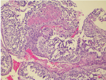
Figure 1: Hemangiopericytoma: Tissue section showed a high cellularity mass with uniform tumor cells with minimal pleomorphism, spindle to round to oval nuclei with vesicular to hyperchromatic chromatin and eosinophilic cytoplasm with indistinct cell borders and richly vascularized with staghorn-appearing vessels, with high mitotic activity. Hematoxylin and Eosin x 40X.
Lupinepublishers-openaccess-Research-Reviews
The histopathologic examination showed a high cellularity mass with uniform tumor cells with minimal pleomorphism, spindle to round to oval nuclei with vesicular to hyperchromatic chromatin and eosinophilic cytoplasm with indistinct cell borders. The tumor was richly vascularized with staghorn-appearing vessels, with high mitotic activity (Figure 1). There was no evidence of tumor tissue infiltrating the bone and the cartilaginous tissues. On immunohistochemistry, the tumor cells were diffusely positive for CD34 and MIC-2 whereas staining for actin and EMA was negative. Our patient is doing well after 12 months of follow up.
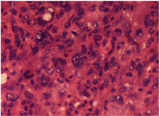
Figure 2: Malignant fibrous histiocytoma: Microscopically, the lesion showed marked architectural and cytologic pleomorphism with haphazardly arranged malignant plump to spindle shaped cells admixed with giant cells. Hematoxylin and Eosin x 40X.
Lupinepublishers-openaccess-Research-Reviews
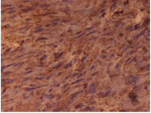
Figure 3: Malignant fibrous histiocytoma: The neoplastic cells were positive for vimentin. IHC Vimentin x 40X.
Lupinepublishers-openaccess-Research-Reviews
Case 2: Pleomorphic Malignant Fibrous Histiocytoma (Mfh): A 63-year-old man presented with a thigh mass for 10 months. On local examination, the mass was 12x 9cm, firm in consistency with ill-defined margins. The specimen of emergent debridement was submitted for pathologic and bacteriologic examination. Microscopically, the lesion showed marked architectural and cytologic pleomorphism with haphazardly arranged malignant plump to spindle shaped cells admixed with giant cells (Figure 2). The neoplastic cells were positive for vimentin (Figure 3), but negative for all lineage-specific markers. The diagnosis of pleomorphic MFH was made. Thoracic computed tomography scan showed bilateral multiple pulmonary nodules. The patient died 1 month later.
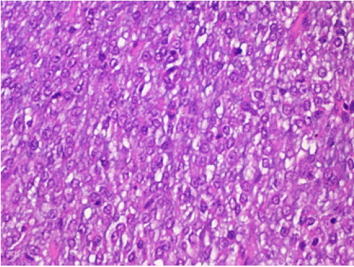
Case 3: Myxoid Liposarcoma: A 25-year-old man presented to the Surgical Clinic with complaints of soft tissue mass in the left thigh for the last 3 months. On local examination, the mass was soft to firm in consistency, well circumscribed of 12x10cm size. Blood parameters were normal and there was no functional abnormality. He was operated upon with wide local excision. The excised mass was 10x9.5cm, soft to gelatinous in consistency without necrosis or haemorrhage. Histopathological examination revealed a myxoid tumor comprising of small dark oval cells in a myxoid background. An extensive capillary network with typical lipoblasts were also seen with mitotic activity of 5 mitosis/ 10HPF (Figure 4). A diagnosis of myxoid liposarcoma was given. The initial surgery was accompanied with adjuvant chemotherapy and complementary radiotherapy. The patient was well after 12 months of follow up.
Figure 4: Myxoid liposarcoma: Histopathological examination showed a myxoid tumor comprising of small dark oval cells in a myxoid background. An extensive capillary network with typical lipoblasts were also seen with mitotic activity of 5 mitosis/ 10HPF. Hematoxylin and Eosin x 40X.
Lupinepublishers-openaccess-Research-Reviews
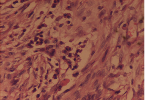
Case 4: Inflammatory Fibrosarcoma: A 42 years old male presented to the Surgery Out-patients Department with complaints of generalised severe pain in the abdomen, localized mainly in the right iliac fossa for 2 days. He was previously fit and healthy and started with generalised feeling of being unwell for four months with mild recurrent pain abdomen, bloating sensation, loss of weight over half a stone and loss of appetite. There was no history of bladder or bowel disturbance, fever, jaundice or vomiting. His pain was worse in the last 2 days associated with nausea. On clinical examination he appeared anxious and sweaty with mild dehydration. Abdominal examination showed tenderness and guarding in the right iliac fossa with the rest of the abdomen soft and there were no masses palpable. Rectal examination was unremarkable. A possible diagnosis of appendicitis was made. Haematological investigations showed a raised white cell count of 17.800/cc and biochemical investigations were within the normal limits. Chest X-Ray was normal with the abdomen plain film showing stones in the gall bladder and a few dilated small bowel loops. Urgent ultrasound scan of abdomen confirmed a small collection of fluid in the right iliac fossa and gallstones with no gas in the biliary tree. In view of the uncertain diagnosis a laparotomy was carried out. This revealed a large mass in the right iliac fossa at the Ileo-caecal junction. The distal ileum was adhered to caecum and adjacent mesentery with thick pus between the loops of small bowel. Right hemicolectomy and an end to side ileo-transverse anastomosis was performed. The postoperative recovery of the patient was uneventful. The histopathology was initially reported as highly cellular spindle cell tumour, with frequent mitotic activity with the most likely site of origin of tumour being bowel wall and spreading in to the surrounding areas including the mesentery. Tumour markers SMA, desmin and vimentin were positive there by indicating leiomyosarcoma. In view of the positive cytokeratin immunostaining, a second opinion was sought by pathologists. On further review, an “Inflammatory fibrosarcoma of the colon” was given due to the marked pleomophism and associated inflammatory cell infiltrate (Figure 5).
Figure 5: Inflammatory Fibrosarcoma: The histopathology revealed a cellular spindle cell tumour, with frequent mitotic activity with marked pleomophism and associated inflammatory cell infiltrate. Hematoxylin and Eosin x 40X.
Lupinepublishers-openaccess-Research-Reviews

Discussion

Hemangiopericytoma is a rare tumor of adult, found mainly in the fifth decade of life. It is most frequently mimicked by synovial sarcoma which occasionally presents as a pure hemangiopericytoma like lesion. These tumors can originate anywhere in the body where there are capillaries. The most common locations reported are the brain, lower extremities, pelvis and head & neck. It originates in the pericytes, the cells normally arranged along specific types of blood vessels. It can be broadly classified as intracranial and extracranial. Extracranially, it can occur at any site throughout the body in soft tissues and bone. They have a grade 2 or 3 behavior and needs to be distinguished from benign meningiomas because of their high rate of recurrence (41%) and metastases (12-20%) [6]. They are highly cellular and mitotically active neoplasm that is rich in pericellular reticulin and stains with anti-type IV collagen. They can be distinguished from benign meningiomas by their hypercellularity, higher mitotic index and microscopically bulge into vascular lumens without bursting through the endothelium, exhibiting a characteristic well-developed “staghorn” branching vascular pattern [7]. They are painless masses and may not have any associated symptoms. They can remain undetected for long periods of time due to the fact that they originate in soft tissue except when intracranial it can cause neurological disturbances. Exhibiting a characteristic well-developed “staghorn” branching vascular pattern. Differential diagnosis includes synovial sarcoma (similar vascular pattern, characteristic translocation), mesenchymal chondrosarcoma (islands of mature cartilage; malignant chondrocytes present), fibrous histiocytoma (storiform pattern, fibrohistiocytic lesion) and solitary fibrous tumor (more prominent collagen, less prominent vessels). Hemangiopericytoma on immunohistochemistry stain positive for CD99, vimentin, CD 34 and negative for Factor VIII and CD31. In adults, complete surgical resection remains the mainstay of treatment. Malignant fibrous histiocytoma has more recently been classified as pleomorphic undifferentiated sarcoma (PUS). It is considered as the most common type of soft tissue sarcoma in adults and has an aggressive biological behaviour with poor prognosis [8,9]. Typically occurs in adults with a slight male predilection. The presentation is usually with a painless, enlarging and well circumscribed palpable mass. They are usually confined to the soft tissues having predilection for extremities but occasionally may arise in or from bone also (1-5%). Some of its histological subtypes includes storiformpleomorphic, myxoid, myxofibrosarcoma, inflammatory, giant cell and angiomatoid. Pleomorphic malignant fibrous histiocytoma which is the most common subtype is considered a diagnosis of exclusion for sarcomas that cannot be more precisely categorized [10]. Imaging typically shows a well-circumscribed mass that is dark on T1-weighted images and bright on T2-weighted images. Histomorphology is characterized by high cellularity, marked nuclear pleomorphism accompanied by abundant mitotic activity including atypical mitoses and a spindle cell morphology. Necrosis is commonly present. Treatment consists of surgical excision and in almost all cases radiation eliminates the need for limb amputation.
Liposarcoma is a malignant tumor that arises from deep soft tissue fat and not from common lipomas. It accounts for up to 20% of all soft tissue sarcomas and commonly affects adult [11]. It can occur in almost any part of the body, commonly involving thigh and retroperitoneum. There are four subtypes, each having its unique characteristics: well-differentiated liposarcoma (most common subtype), myxoid/round cell liposarcoma, pleomorphic liposarcoma(rarest subtype) and dedifferentiated liposarcoma. Dedifferentiated liposarcoma and pleomorphic liposarcoma are considered as highly malignant [12]. Myxoid liposarcoma (MLS) is considered as a low grade tumor but the presence of areas of round cells more than 5% is associated with a worse prognosis [4]. MLS presents as a slow-growing, deep-seated tumor in the lower extremity of a relatively young adult. Specific chromosomal translocations have been discovered in MLS which consists of the fusion of the FUS and CHOP genes [(t12;16)(q13;p11)] in 90% of tumor [13]. Extremity myxoid liposarcomas have an unusually high predilection for extra-pulmonary metastases often deep soft tissue locations such as retroperitoneum or extrimities without any pulmonary metastases [14,15]. Imaging of the abdomen, retroperitoneum, and extrapleural chest should be performed for accurate staging and post-treatment follow-up of patients with myxoid liposarcoma. In all patients surgical management of the tumor is curative. It is radiosensitive as compared with other soft tissue sarcomas [16]. After treatment of the primary tumor, such patients should be followed with regular chest X-ray and abdominal/pelvic computed tomography (CT) scans.
Fibrosarcoma is a malignant neoplasm of mesenchymal origin in which histologically the predominant cells are fibroblasts that divide excessively without cellular control. Inflammatory fibrosarcoma is rare and was originally described in the lung by names such as pseudotumor, inflammatory pseudotumor and plasma cell granuloma. It is typically considered as a benign tumor with aggressive behavior (low-grade tumor) that can occur anywhere in the body. They are solitary, well-demarcated fibrous tumors and are characterized by storiform pattern of fibrous tissue along with of mixture of inflammatory cells such as plasma cells, lymphocytes and eosinophils as well as spindle cells without nuclear atypia [17]. These tumors may also have necrosis, hemorrhage, focal calcification and mitotic activity. Some of the histologic differential diagnosis includes: calcifying fibrous pseudotumor, inflammatory fibroid tumor and nodular fasciitis. Immunohistochemically, the tumor shows intense immunoreactivity for vimentin, muscle actin, and α-smooth muscle actin but are negative for desmin and highmolecular- weight caldesmon. In addition, tumor cells are not labeled by antibodies against AE1/3 and CAM5.2. The prognosis is generally good on tumor removal, but rarely some tumors are known to metastasize.

For more Lupine Publishers Open Access Journals Please visit our website:
http://lupinepublishers.us/
For more Research and Reviews on Healthcare articles Please Click Here:
https://lupinepublishers.com/research-and-reviews-journal/
To Know More About Open Access Publishers Please Click on Lupine Publishers
Follow on Linkedin : https://www.linkedin.com/company/lupinepublishers
Follow on Twitter   :  https://twitter.com/lupine_online


Lupine Publishers: Lupine Publishers| A Standard Pediatric Dental Clinic

Lupine Publishers: Lupine Publishers| A Standard Pediatric Dental Clinic :  Lupine Publishers| Journal of Dentistry and Oral Health Care Aft...